
Seminário de Seminário de Física e de Engenharia Biomédica
Título: Image guided proton therapy for the treatment of cancers
Orador: Rudi Labarbe (IBA, Université de Liège, Belgium)
Zoom: https://videoconf-colibri.zoom.us/j/84718988078
Abstract:
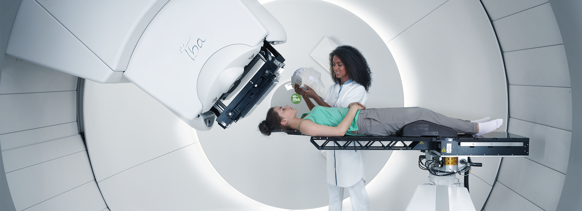
Proton therapy is considered the most advanced form of radiation therapy that uses high-energy proton beam to irradiate tumors. Proton therapy is used today to treat many cancers and is particularly appropriate in situations where treatment options are limited and conventional radiotherapy using photon beam presents unacceptable risks to patients. These situations include eye and brain cancers, head and neck cancers, prostate, liver, lung, breast, and pediatric cancers, as well as other tumors in close proximity to one or more critical structures. Protons, accelerated to therapeutic energies ranging from 70 to 250 MeV, typically with a cyclotron, are transported to the treatment room where they enter the treatment head mounted on a rotating gantry. Spreading and shaping can be achieved by using magnetic scanning of thin “beamlets” of protons of a sequence of initial energies. The latter technique can be used to treat patients with optimized intensity modulated proton therapy (IMPT), the most powerful proton modality. In the treatment of cancer, protons exhibit advantageous dose deposition properties with a low dose plateau followed by a sharp raise of the dose at the end of their range (Bragg peak).
A drawback of using protons over photons is the sensitivity of proton to anatomical changes that translates into uncertainties in the proton range. In lungs, this problem is magnified by continuous anatomical deformations due to breathing. Therefore, having real-time information about tumor position and its surrounding anatomy is of great value in treating lung cancer with proton therapy. At the beginning of a treatment fraction, a Cone Beam Computed Tomography (CBCT) acquisition is performed, and used to reposition the patient. Obtaining a 4D reconstruction from this cone beam data would allow the therapists to check whether the breathing motion of the day still matches that of the planning CT, and if not, take appropriate corrective actions. But 4D tomography from a single cone beam CT acquisition implies a severe lack of projection data, and efficient methods have only started to appear during the last few years. Some perform regularization along time to explicitly enforce similarity between consecutive frames, which considerably improves image quality. In Image Guided Radiotherapy (IGRT), breathing motion can be estimated on the 4D planning CT, and used to refine the 4D CBCT reconstruction results.